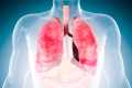
Scientists Discover Exactly How COVID-19 Wreaks Havoc on Human Lungs
0 View

- Publish Date:
- 8 June, 2021
- Category:
- Covid
- Video License
- Standard License
- Imported From:
- Youtube
New structure shows how virus envelope protein hijacks cell junction protein and promotes viral spread; findings could accelerate drug design to block severe effects of COVID-19.
Scientists at the US Department of Energy’s (DOE) Brookhaven National Laboratory have published the first detailed atomic-level model of the SARS-CoV-2 ‘envelope’ protein bound to a human protein essential for the maintenance of the lining of the lungs. The model showing how the two proteins interact, just published in the journal Nature Communications, helps explain how the virus can cause extensive lung damage and escape the lungs to infect other organs in particularly vulnerable COVID-19 patients. The findings may accelerate the search for drugs to block the most serious effects of the disease.
“By obtaining atomic-level details of the protein interactions, we can explain why the damage occurs and look for inhibitors that can specifically block these interactions,” said lead author Qun Liu, a structural biologist at Brookhaven Lab. “If we can find inhibitors, the virus won’t do nearly as much damage. That could give people with compromised health a much better chance for their immune systems to successfully fight the virus.”
New structure shows how the envelope protein of the COVID-19 virus (E, magenta sticks) interacts with a human cell junction protein (PALS1, surfaces colored in blue, green and orange). Understanding this complex structure, which was solved using a cryoelectron microscope at Brookhaven National Laboratory, could lead to the discovery of drugs that block the interaction and possibly the most severe effects of COVID-19. Credit: Brookhaven National Laboratory
Scientists uncovered the details and developed the molecular model using one of the new cryoelectron microscopes at Brookhaven Lab’s Laboratory for BioMolecular Structure (LBMS), a new research facility built with funding from the State of New York adjacent to Brookhaven’s National Synchrotron Light Source II (NSLS). -II).
“LBMS opened ahead of schedule last summer because of its importance in the fight against COVID-19,” said Sean McSweeney, director of LBMS and co-author of the paper. “LBMS and NSLS-II provide complementary protein imaging techniques and both play an important role in deciphering the details of proteins involved in COVID-19. This is the first article published based on the results of the new facility.”
Liguo Wang, scientific director of LBMS and another co-author of the paper, explained that “cryoelectron microscopy (cryo-EM) is particularly useful for studying membrane proteins and dynamic protein complexes, which can be difficult to crystallize for protein crystallography, another common technique for studying protein structures.With this technique we created a 3D map from which we could see how the individual protein components fit together.”
“Without cryo-EM, we wouldn’t have been able to get a structure to capture the dynamic interactions between these proteins,” Liu said.
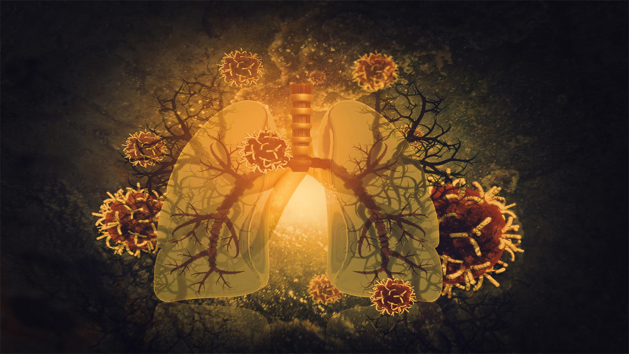
Cause lung disturbance
The SARS-CoV-2 envelope protein (E), found on the outer membrane of the virus alongside the now infamous coronavirus spike protein, helps assemble new virus particles in infected cells. Studies published early in the COVID-19 pandemic showed that it also plays a critical role in hijacking human proteins to facilitate the delivery and transmission of viruses. Scientists hypothesize it does this by binding to human cell junction proteins, pulling them away from their usual job of keeping the connections between lung cells tightly sealed.
“That interaction could be good for the virus and really bad for people — especially older COVID-19 patients and those with pre-existing medical conditions,” Liu said.
A close-up view of the envelope protein of the COVID-19 virus (magenta) and its interaction with specific amino acids that form a hydrophobic pocket on PALS1 (blue, green, and orange). Credit: Brookhaven National Laboratory
When lung cell connections are disrupted, immune cells come in to try to repair the damage, releasing small proteins called cytokines. This immune response can make matters worse by triggering massive inflammation, which triggers a so-called “cytokine storm” and subsequent acute respiratory distress syndrome.
Because the damage weakens cell-cell connections, it can also make it easier for the viruses to escape from the lungs and travel through the bloodstream to infect other organs, including the liver, kidneys and blood vessels.
“In this scenario, the most damage would occur in patients with more viruses and more E proteins being produced,” Liu said. And this could become a vicious cycle: more viruses making more E proteins and more cell junction proteins being pulled back, causing more damage, more transmission, and more viruses again. In addition, existing damage, such as scarring in lung cells, would likely make it more difficult for COVID patients to recover from the damage.
“So we wanted to study this interaction — to understand the atomic-level details of how E interacts with one of these human proteins to learn how to interrupt the interactions and reduce or block these severe effects,” Liu said.
From dots to blobs to card to model
The scientists obtained atomic-level details of the interaction between E and a human lung-cell junction protein called PALS1 by mixing the two proteins together, rapidly freezing the sample, and then studying the frozen sample with the cryo-EM. The electron microscopes use high-energy electrons to interact with the sample in much the same way that ordinary light microscopes use light rays. But electrons allow scientists to see things on a much smaller scale because of their extremely short wavelength (100,000 times shorter than that of visible light).
The first images seemed little more than specks. But image-processing techniques allowed the team to select specks that were true complexes of the two proteins.
Deciphering the structure of the COVID-19 virus E protein bound to human PALS1: starting with a motion-corrected cryo-EM micrograph of nanometer-scale granular dots (a), image processing and two-dimensional averaging produced low-resolution projections of the bound proteins from different orientations (b). Computer tools then transformed these 2D images into a 3D map (c). Blue indicates the most stable parts with the highest resolution and red indicates parts with lower resolution with more flexibility. This map provides enough detail to fit the amino acid building blocks of the two proteins into a final structure of the complex (d), where different parts of PALS1 are shown in blue, green and orange and the viral E protein is magenta. Credit: Brookhaven National Laboratory
“We used two-dimensional averaging and started to see some structural features shared by these particles. Our images showed the complex from different orientations, but at a rather low resolution,” Liu said. “We then use computational tools and infrastructure at Brookhaven’s Computational Science Initiative to perform three-dimensional reconstructions. These give us a 3D model – an experimental map of the structure.”
With an overall resolution of 3.65 Angstroms (the size of just a few atoms), the map contained enough information about the unique characteristics of the individual amino acids that make up the two proteins for the scientists to piece together the known structures of those amino acids. to fit. the card.
“We can see how the chain of amino acids that make up the PALS1 protein folds to form three structural components or domains, and how the much smaller chain of amino acids that make up the E protein folds into a hydrophobic pocket between two of those two.” fits. domains,” said Liu.
The model provides both the structural details and an understanding of the intermolecular forces that allow E proteins to wrench PALS1 from its place deep within an infected cell at the cell’s outer boundary.
“Now we can explain how the interactions pull PALS1 out of the human lung cell junction and contribute to the damage,” Liu said.
Consequences for drugs and evolution
“This structure provides the basis for our computational science colleagues to conduct docking studies and molecular dynamic simulations to look for drugs or drug-like molecules that can block the interaction,” said John Shanklin, chair of Brookhaven Lab’s Department of Biology and a co-author of the study. author of the paper. “And if they identify promising leads, we have the analytical capabilities to rapidly screen such drug candidates to identify those that may hold the key to preventing severe impacts from COVID-19.”
By understanding the dynamics of this protein interaction, scientists can also track how viruses like SARS-CoV-2 evolve.
“If the virus protein PALS1 pulls out of the cell junction, it can help to spread the virus more easily. That would give a selective advantage to the virus. Any traits that increase the survival, spread or delivery of the virus are likely to be preserved,” Liu said.
The longer the virus continues to circulate, the more likely it is that new evolutionary advantages will emerge.
“This is another reason why it is so vital for us to identify and implement promising therapies,” Liu said. “In addition to preventing the most serious infections, drugs that effectively treat COVID-19 will keep us ahead of these mutations.”
Reference: June 8, 2021, Nature Communications.
DOI: 10.1038/s41467-021-23533-x
This research was funded by Brookhaven National Laboratory’s COVID-19 Laboratory Directed Research and Development (LDRD) fund. LBMS is supported by the DOE Office of Science (BER), NSLS-II is a DOE Office of Science user facility, supported by the Office of Science (BES).