New Research Shows COVID-19 Alters Gray Matter Volume in the Brain
0 View

- Publish Date:
- 12 May, 2021
- Category:
- Covid
- Video License
- Standard License
- Imported From:
- Youtube
Covid-19 patients who receive oxygen therapy or experience a fever show decreased gray matter volume in the brain’s frontal-temporal network, according to a new study led by researchers at Georgia State University and the Georgia Institute of Technology.
The study found that a lower gray matter volume in this brain region was associated with a higher level of disability in Covid-19 patients, even six months after hospital discharge.
Gray matter is vital for information processing in the brain, and abnormal gray matter can affect how well neurons function and communicate. The study, published in the May 2021 issue of Neurobiology of Stress, indicates that frontal network gray matter could be a core area for brain involvement in Covid-19, even beyond damage related to clinical manifestations of the disease, such as a stroke.
The researchers, who are affiliated with the Center for Translational Research in Neuroimaging and Data Science (TReNDS), analyzed computed tomography scans in 120 neurological patients, including 58 with acute Covid-19 and 62 without Covid-19, matched for age, gender, and disease. The work was carried out together with Enrico Premi and his colleagues from the University of Brescia in Italy, who provided the data for the study. They used source-based morphometry analysis, which increases statistical power for studies with a moderate sample size.
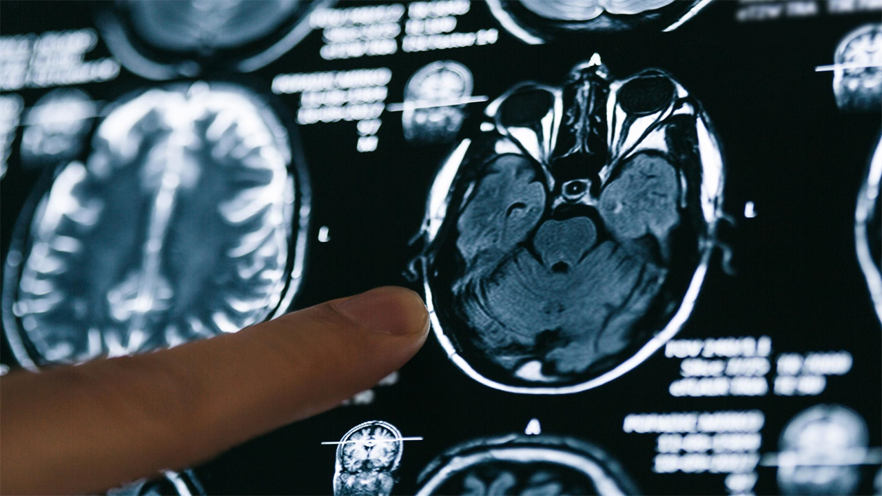
Researchers Kuaikuai Duan and Vince Calhoun have found that neurological complications in Covid-19 patients may be related to a lower volume of gray matter in the front part of the brain, even six months after discharge from the hospital. Credit: Vince Calhoun, Georgia Tech
“Science has shown that brain structure influences its function, and abnormal brain imaging has emerged as an important feature of Covid-19,” said Kuaikuai Duan, lead author of the study, a graduate research assistant. at TReNDS and Ph.D. student at Georgia Tech’s School of Electrical and Computer Engineering. “Previous studies have examined how the brain is affected by Covid-19 using a univariate approach, but ours is the first to use a multivariate, data-driven approach to link these changes to specific Covid-19 characteristics (for example, fever). and lack of oxygen) and outcome (disability level). “
The analysis showed that patients with higher disability had lower gray matter volume in the superior, medial, and middle frontal gyri at discharge and six months later, even at control for cerebrovascular disease. The gray matter volume in this region was also significantly reduced in patients who received oxygen therapy compared to patients who did not receive oxygen therapy. Febrile patients had a significant decrease in gray matter volume in the inferior and middle temporal gyri and spindle-shaped gyrus compared to patients without fever. The results suggest that Covid-19 may affect the frontal-temporal network through fever or oxygen starvation.
Reduced gray matter in the superior, medial, and middle frontal gyri was also present in patients with agitation compared to patients without agitation. This implies that changes in gray matter in the frontal region of the brain may underlie the mood disorders that commonly occur in Covid-19 patients.
“Neurological complications are increasingly being documented for patients with Covid-19,” said Vince Calhoun, senior author of the study and director of TReNDS. Calhoun is a Distinguished University Professor of Psychology at Georgia State and holds positions in the School of Electrical and Computer Engineering at Georgia Tech and in Neurology and Psychiatry at Emory University. “Gray matter reduction has also been shown to be present in other mood disorders such as schizophrenia and is likely related to the way gray matter affects neuron function.”
The study’s findings indicate that changes in the frontal-temporal network can be used as a biomarker to determine the likely prognosis of Covid-19 or to evaluate treatment options for the disease. Next, the researchers hope to replicate the study on a larger sample size that includes many types of brain scans and different populations of Covid-19 patients.
Reference: “Frontal-temporal gray matter volume changes associated with clinical measurements of older adults with COVID-19” by Kuaikuai Duan, Enrico Premi, Andrea Pilotto, Viviana Cristillo, Alberto Benussi, Ilenia Libri, Marcello Giunta, H. Jeremy Bockholt, Jingyu Liu, Riccardo Campora, Alessandro Pezzini, Roberto Gasparotti, Mauro Magoni, Alessandro Padovani, and Vince D. Calhoun, Apr 13, 2021, Neurobiology of Stress.
DOI: 10.1016 / j.ynstr.2021.100326
TReNDS is a partnership between Georgia State, Georgia Tech and Emory University and aims to improve our understanding of the human brain through advanced analytical approaches. The center uses large-scale data exchange and multimodal data fusion techniques, including deep learning, genomics, brain mapping and artificial intelligence.