New Cell Atlas of COVID-19 Pathology Reveals How the Coronavirus “Wreaks Havoc in the Lungs”
0 View

- Publish Date:
- 13 June, 2021
- Category:
- Covid
- Video License
- Standard License
- Imported From:
- Youtube
Single-cell analysis of autopsy samples from COVID-19 patients shows how the lungs repeatedly tried, and failed, to repair themselves.
Scientists from several hospitals and research centers have shown what happens in individual cells of patients who have died from COVID-19. In a study published in Nature, the researchers describe how infected cells from multiple organs showed a range of molecular and genomic changes. They also saw signs of multiple, unsuccessful attempts by the lungs to repair itself in response to respiratory failure, the leading cause of death in COVID-19 patients.
“You really feel the tragedy of the disease when you see that result,” said Aviv Regev, co-senior author of the study and a core member of the Broad Institute at MIT and Harvard when the study began. “The lung is trying everything it has at its disposal, and it still can’t repair itself. This was a very emotional study. We are grateful to the patients and families who have agreed to donate tissue for COVID-19 research to advance our understanding of this devastating disease.”
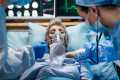
The researchers studied tissue obtained from autopsies of 17 individuals who succumbed to COVID-19 and were cared for at Beth Israel Deaconess Medical Center, Brigham and Women’s Hospital and Massachusetts General Hospital.
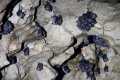
Researchers profiled lung tissue from deceased COVID-19 patients and zoomed in on key regions and structures of interest. Credit: Domenic Abbondanza
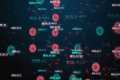
The team investigated how the SARS-CoV-2 virus disrupts the function of cells and their genetic programs. They used single-cell RNA sequencing data from tissue samples taken from 11 organ systems — including the lungs, heart, liver and kidneys — to build a comprehensive “cell atlas” of hundreds of thousands of individual cells showing how COVID-19 could lead to organ failure and death.
“We knew people were dying from COVID-related pneumonia and extrapulmonary complications,” said Alexandra-Chloé Villani, an associate member of the Broad, a principal investigator at Mass General, an assistant professor of medicine at Harvard Medical School, and co-senior. author of the study. “Before this study, we had limited knowledge of the cellular and molecular mechanisms involved in a patient’s death.”
The study describes the results of a collaboration of researchers from the Broad Institute, Mass General, the Ragon Institute of MGH, MIT and Harvard, MIT, Beth Israel Deaconess Medical Center, Brigham and Women’s Hospital, Columbia University Irving Medical Center and other institutions. A team led by the Columbia collaborators co-authored an accompanying study also published in Nature.
The team’s cell atlas is freely and openly available for other scientists to explore. They also created a biobank with 420 copies of the autopsy samples that can be used for other COVID-19 studies. “We’ve created a foundational resource that other researchers can use in the future to ask specific questions,” said Orit Rozenblatt-Rosen, co-senior author and an institute scientist and the scientific director of the Klarman Cell Observatory at the Broad when the study was conducted. began . “Hopefully our findings will enable people to find better therapies for COVID-19.”
New techniques for a new virus
To learn more about cellular mechanisms underlying organ failure caused by COVID-19, the researchers knew they had to study the organs themselves. For that they would need samples from autopsies.
Working with autopsy samples is challenging under normal circumstances. To treat samples that may contain a novel, highly contagious pathogen, the researchers developed novel tissue collection and processing protocols compatible with biosafety level 3 laboratory requirements.
“We wanted to make sure we could learn and share as much as possible to help prevent future deaths, while prioritizing the safety and well-being of everyone involved. This was no small feat, given the COVID-related limitations and all the uncertainties surrounding it. It was amazing to watch dozens of scientists and medical professionals from different institutes come together as a partnership to carefully design and coordinate our experimental and computational efforts,” said institute member and co-senior author Alex K. Shalek, who is also a member of the Ragon Institute, and an associate professor of chemistry, a core member of the Institute for Medical Engineering and Science, and an extramural member of the Koch Institute for Integrative Cancer Research at MIT.
The team then profiled RNA from the individual cells and developed new methods to analyze and annotate the large amounts of sequence data. They compared gene expression signatures of different cells: COVID-19-damaged cells and uninfected cells from the COVID-19 patients, as well as cells from patients with other diseases and from healthy individuals.
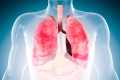
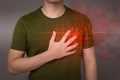
Havoc in the lungs
The most comprehensive set of findings came from the lungs. The scientists were amazed at the magnitude of the changes in genetic programs they found there. “The virus is wreaking havoc in the lungs and we see it in the cells,” Regev said.
A major cause of lung damage in COVID-19 is the destruction of AT1 cells, which enable respiration and gas transfer. The scientists found that when AT1 cells died, related lung cells, called AT2, tried to convert themselves into AT1 cells through a process called transdifferentiation. But this attempt stopped halfway through, leaving the cells in an intermediate state often seen in patients with other lung diseases such as pulmonary fibrosis.
In a final attempt at self-repair, the lungs attempted to convert cells from higher up in the airways known as intrapulmonary basal-like progenitor cells into AT1 cells. This attempt at transdifferentiation had only been seen before in mouse models.
The findings suggest that the lung failure in patients was caused by the inability of lung cells to surpass the damage caused by the virus as the cells tried to regenerate.
Change programs
The article also describes how the virus affects other tissues outside the lungs. A surprising finding was that, although the heart suffered significant damage and showed evidence of altered genetic programs in many different cell types, there was very little viral RNA in the heart tissue itself. “Whether that means the virus had already cleared, or the heart was collateral damage is an area for further investigation,” Regev said.
The researchers also looked at 27 different genes that previous genome-wide association studies have linked to severe COVID-19. They focused on a handful that were highly expressed in the main cell types in the new study, specifically those in infected lungs. This finding helps refine the list of potential genetic factors for serious diseases and highlights the cell types that may be most relevant in severe COVID-19.
The team now plans to complete the analysis of the other autopsy tissues, such as brain, spleen and trachea, to paint a more complete picture of COVID-19 pathology and provide a resource for future studies. .
To learn more about this research, read New Cell Atlas of COVID Lungs Reveals Why SARS-CoV-2 Is Different and Deadly.
Reference: “COVID-19 Tissue Atlases Reveal SARS-CoV-2 Pathology and Cellular Targets” by Toni M. Delorey, Carly GK Ziegler, Graham Heimberg, Rachelly Normand, Yiming Yang, sa Segerstolpe, Domenic Abbondanza, Stephen J. Fleming, Ayshwarya Subramanian, Daniel T. Montoro, Karthik A. Jagadeesh, Kushal K. Dey, Pritha Sen, Michal Slyper, Yered H. Pita-Juárez, Devan Phillips, Jana Biermann, Zohar Bloom-Ackermann, Nikolaos Barkas, Andrea Ganna, James Gomez , Johannes C. Melms, Igor Katsyv, Erica Normandin, Pourya Naderi, Yury V. Popov, Siddharth S. Raju, Sebastian Niezen, Linus T.-Y. Tsai, Katherine J. Siddle, Malika Sud, Victoria M. Tran, Shamsudheen K. Vellarikkal, Yiping Wang, Liat Amir-Zilberstein, Deepak S. Atri, Joseph Beechem, Olga R. Brook, Jonathan Chen, Prajan Divakar, Phylicia Dorseus, Jesse M. Engreitz, Adam Essene, Donna M. Fitzgerald, Robin Fropf, Steven Gazal, Joshua Gould, John Grzyb, Tyler Harvey, Jonathan Hecht, Tyler Hether, Judit Jané-Valbuena, Michael Leney-Greene, Hui Ma, Cristin McCabe, Daniel E. McLoughlin, Eric M. Miller, Christoph Muus, Mari Niemi, Robert Padera, Liuliu Pan, Deepti Pant, Carmel Pe’er, Jenna Pfiffner-Borges, Christopher J. Pinto, Jacob Plaisted, Jason Reeves, Marty Ross, Melissa Rudy, Erroll H. Rueckert, Michelle Siciliano, Alexander Sturm, Ellen Todres, Avinash Waghray, Sarah Warren, Shuting Zhang, Daniel R. Zollinger, Lisa Cosimi, Rajat M. Gupta, Nir Hacohen, Hanina Hibshoosh, Winston Hide, Alkes L. Price, Jayaraj Rajagopal, Purushothama Rao Tata, Stefan Riedel, Gyongyi Szabo, Timothy L. Tickle, Patrick T. Ellinor, Debora h Hung, Pardis C. Sabeti, Richard Novak, Robert Rogers, Donald E. Ingber, Z. Gordon Jiang, Dejan Juric, Mehrtash Babadi, Samouil L. Farhi, Benjamin Izar, James R. Stone, Ioannis S. Vlachos, Isaac H Solomon, Orr Ashenberg, Caroline BM Porter, Bo Li, Alex K. Shalek, Alexandra-Chloé Villani, Orit Rozenblatt-Rosen, and Aviv Regev, April 29, 2021, Nature.
DOI: 10.1038/s41586-021-03570-8
Aviv Regev is now Executive Vice President, Genentech Research and Early Development.
Orit Rozenblatt-Rosen is now Executive Director and Head of Cellular and Tissue Genomics at Genentech.
Support for this research was provided in part by the Manton Foundation, Klarman Family Foundation, Howard Hughes Medical Institute, the Chan Zuckerberg Initiative and the trans-network projects of the Human Tumor Atlas Network SARDANA (Shared Repositories, Data, Analysis and Access), DARPA, and the US Food and Drug Administration.