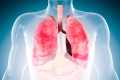
New Cell Atlas of COVID Lungs Reveals Why SARS-CoV-2 Is Different and Deadly
0 View

- Publish Date:
- 2 May, 2021
- Category:
- Covid
- Video License
- Standard License
- Imported From:
- Youtube
A new study paints the most detailed picture to date of SARS-CoV-2 infection in the lungs, revealing mechanisms that lead to lethal COVID-19, potentially explaining long-term complications and showing how COVID -19 differs from other infectious diseases.
Led by researchers at Columbia University Vagelos College of Physicians and Surgeons and Herbert Irving Comprehensive Cancer Center, the study found that in patients who died of the infection, COVID-19 unleashed a damaging trifecta of runaway inflammation, direct destruction, and reduced lung cell regeneration. involved in gas exchange and accelerated lung scarring.
Although the study looked at the lungs of patients who had died of the disease, it provides solid evidence as to why survivors of severe COVID may experience long-term respiratory complications due to lung scarring.
“It’s a devastating disease, but the picture we are getting of the COVID-19 lung is the first step towards identifying potential targets and therapies that disrupt some of the disease’s vicious circuitry. In particular, targeting cells responsible for pulmonary fibrosis early on could potentially prevent or improve long-term complications in survivors of severe COVID-19, ”said Benjamin Izar, MD, PhD, assistant professor of medicine, who led to a group of more than 40 researchers to complete a series of analyzes in a few months that usually takes years.
This study and an accompanying article led by Harvard / MIT researchers, to which the Columbia researchers also contributed, were published April 29 in the journal Nature.
Study makes atlas of cells in COVID lung
The new study is unique from other studies in that it directly examines lung tissue (rather than sputum or bronchial washes) using single-cell molecular profiling that can identify each cell in a tissue sample and record each cell’s activity, resulting in an atlas of cells. in COVID lung.
“A normal lung will have many of the same cells that we find in COVID, but in different ratios and different activation states,” says Izar. “To understand how COVID-19 differs from both control lungs and other forms of infectious pneumonia, we had to look at thousands of cells one at a time.”
Izar’s team examined the lungs of 19 individuals who died of COVID-19 and underwent a rapid autopsy (within hours of death) – during which lung and other tissue were collected and immediately frozen – and the lungs of non-COVID- 19 patients. In collaboration with researchers at Cornell University, the researchers also compared their findings with lungs of patients with other respiratory diseases.
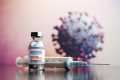
Drugs targeting IL-1ß can reduce inflammation
Compared to normal lungs, the COVID patients’ lungs were filled with immune cells called macrophages, the study found.
Typically during an infection, these cells chew up pathogens, but also regulate the intensity of the inflammation, which also aids in the fight.
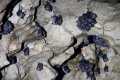
The lungs of patients with COVID-19 have more monocytes expressing IL-1beta than lungs of patients with other respiratory conditions. Credit: Benjamin Izar.
“In COVID-19 we see expansion and uncontrolled activation of macrophages, including alveolar macrophages and monocyte-derived macrophages,” says Izar. “They are completely out of balance and increase inflammation uncontrollably. This results in a vicious cycle in which more immune cells come in and cause even more inflammation, ultimately damaging the lung tissue. “
In particular, one inflammatory cytokine, IL-1β, is highly produced by these macrophages.
“In contrast to other cytokines such as IL-6, which appear to be universal in various pneumonia, IL-1β production in macrophages is more pronounced in COVID-19 compared to other viral or bacterial lung infections,” says Izar. “This is important because there are drugs that reduce the effects of IL-1ß.”
Some of these drugs are already being tested in clinical trials with COVID patients.
Severe COVID also prevents lung recovery
In a typical infection, a virus damages lung cells, the immune system clears the pathogen and debris, and the lung recovers.
But in COVID, the new study found that not only does the SARS-CoV-2 virus destroy alveolar epithelial cells important for gas exchange, the resulting inflammation also impairs the remaining cells’ ability to regenerate the damaged lung.
Lung cells in patients with severe COVID become stuck in a state (indicated by the green color) that prevents the cells from repairing the damage caused by the infection. The left image shows cells from a healthy lung; the right image shows lung cells from a patient who died from COVID-19. Credit: Benjamin Izar / Columbia University Vagelos College of Physicians and Surgeons.
Although the lung still contains cells that can make the repairs, inflammation permanently traps these cells in an intermediate cell state, rendering them unable to complete the final steps of differentiation required for adult lung epithelium replacement.
“IL-1β, among others, appears to be a culprit in inducing and maintaining this intermediate cell state,” says Izar, “associating inflammation and decreased lung regeneration in COVID-19, suggesting that in addition to reducing inflammation , targeting IL-1β can help to reduce the inhibition of the cells needed for lung repair. “
Prevent accelerated fibrosis
The researchers also found a large number of specific fibroblast cells, called pathological fibroblasts, that cause rapid scarring in COVID-19 lungs. When the fibroblast cells fill the lung with scar tissue, a process called fibrosis, the lung has less space for cells involved in gas exchange and is permanently damaged.
Given the importance of pathological fibroblasts in the disease, Izar’s team carefully analyzed the cells to discover potential drug targets. An algorithm called VIPER, previously developed by Andrea Califano, Dr., chair of systems biology at Columbia University Vagelos College of Physicians and Surgeons, identified several molecules in the cells that play important roles and may be targets of existing drugs.
“This analysis predicted that inhibition of STAT signaling could alleviate some of the deleterious effects caused by pathological fibroblasts,” said Izar.
“We hope that by sharing this analysis and huge data sources, other researchers and pharmaceutical companies can begin to test and expand these ideas and find treatments to not only treat critically ill patients, but also to reduce complications in humans. that serious COVID- 19. “
Team effort by several Columbia labs
“Bringing this study together in such a short time was only possible with the help of different teams of researchers in Columbia,” said Izar.
Critically, in the first few months of the pandemic, the Department of Pathology and Cell Biology of Colombia decided to rapidly freeze many tissues from deceased COVID patients to preserve the molecular state of the cells. Hanina Hibshoosh, MD, director of the department’s tissue bank, initiated collaboration with Izar’s laboratory, which has expertise in performing single-cell analyzes on frozen tissue. Pathologist Anjali Saqi, MD, professor of pathology and cell biology, was also instrumental in obtaining and evaluating the samples.
Jianwen Que, MD, PhD, professor of medicine, and his laboratory provided expertise in identifying and characterizing cells in the lung and their regenerative potential. Fibrosis expert Robert Schwabe, MD, associate professor of medicine, was essential in dissecting mechanisms by which COVID-19 caused lung scarring.
“We are incredibly grateful to all the laboratories that contributed to this effort and we are fortunate to be at Columbia with all the necessary expertise in one collaborative environment,” said Izar.
Reference: “A single-cell molecular long atlas of lethal COVID-19” by Johannes C. Melms, Jana Biermann, Huachao Huang, Yiping Wang, Ajay Nair, Somnath Tagore, Igor Katsyv, André F. Rendeiro, Amit Dipak Amin, Denis Schapiro, Chris J. Frangieh, Adrienne M. Luoma, Aveline Filliol, Yinshan Fang, Hiranmayi Ravichandran, Mariano G. Clausi, George A. Alba, Meri Rogava, Sean W. Chen, Patricia Ho, Daniel T. Montoro, Adam E. Kornberg, Arnold S. Han, Mathieu F. Bakhoum, Niroshana Anandasabapathy, Mayte Suárez-Fariñas, Samuel F. Bakhoum, Yaron Bram, Alain Borczuk, Xinzheng V. Guo, Jay H. Lefkowitch, Charles Marboe, Stephen M. Lagana, Armando Del Portillo, Emmanuel Zorn, Glen S. Markowitz, Robert F. Schwabe, Robert E. Schwartz, Olivier Elemento, Anjali Saqi, Hanina Hibshoosh, Jianwen Que and Benjamin Izar, April 29, 2021, Nature.
DOI: 10.1038 / s41586-021-03569-1
The researchers were supported by the US National Institutes of Health (grants K08CA222663, U54CA225088, R37CA258829, R01HL152293, R01HL132996, T32CA203702, UL1TR002384, R01CA194547, R01CA234614, R01AI107301, R01AI107301, R01AI107301, R01AI107301, R01AI107301, R01AI107301, R01AI107301, R01AI107301, R01AI107301, R01AI107301 , R01AI107301); FastGrants; a Burroughs Wellcome Fund Career Award for medical scientists; the Louis V. Gerstner Jr. Scholars Program; the United States Department of Defense (Discovery Award PR200616); Research grants Volastra, Janssen and Eli Lilly; the Leukemia and Lymphoma Society (grants SCOR 7012-16, SCOR 7021-20 and SCOR 180078-02); an Irma Hirschl Trust Research Award Scholar; and the Damon Runyon Cancer Research Foundation (DRQ-03-20).
This research was funded in part through NIH Support Grant S10RR027050 for flow cytometry analysis and NIH / NCI Cancer Center Support Grant P30CA013696 from Columbia University’s Genetically Modified Mouse Model Shared Resource, Molecular Pathology Shared Resource, and Tissue Bank.
Benjamin Izar is a consultant to Merck and Volastra Therapeutics. Olivier Elemento is a scientific advisor and shareholder in Freenome, Owkin, Volastra Therapeutics and OneThree Biotech. Robert E. Schwartz is a member of the Scientific Advisory Board of Miromatrix Inc. Daniel T. Montoro is a consultant for LASE Innovation.