Coronavirus Does Not Infect the Brain, but Still Inflicts Significant Neurological Damage
0 View

- Publish Date:
- 21 April, 2021
- Category:
- Covid
- Video License
- Standard License
- Imported From:
- Youtube
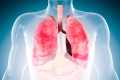
SARS-CoV-2, the virus that causes COVID-19, probably doesn’t directly infect the brain, but it can still cause significant neurological damage, according to a new study by neuropathologists, neurologists and neuroradiologists at Columbia University Vagelos College of Physicians and Surgeons .
“There has been much debate as to whether this virus infects the brain, but we couldn’t find signs of virus in brain cells from more than 40 COVID-19 patients,” said James E. Goldman, MD, PhD, professor of pathology and disease. cell biology (in psychiatry), who led the study with Peter D. Canoll, MD, PhD, professor of pathology and cell biology, and Kiran T. Thakur, MD, the Winifred Mercer Pitkin assistant professor of neurology.
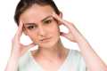
“At the same time, we have observed many pathological changes in this brain, which could explain why critically ill patients experience confusion and delirium and other serious neurological effects – and why people with mild cases may experience ‘brain fog’ for weeks and months.”
The study, published in the journal Brain, is the largest and most detailed COVID-19 brain autopsy report published to date, suggesting that the neurological changes commonly seen in these patients may be due to inflammation caused by the virus in other parts of the body or in the blood vessels of the brain.
No virus in brain cells
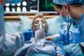
The study examined the brains of 41 patients with COVID-19 who succumbed to the disease during their hospitalization. The patients ranged in age from 38 to 97 years; about half had been intubated and all had lung damage caused by the virus. Many of the patients were of Hispanic ethnicity. There was a wide range of hospital durations, with some patients dying shortly after arriving in the emergency room, while others remaining in the hospital for months. All patients underwent extensive clinical and laboratory examinations, and some had MRI and CT scans of the brain.
To detect any virus in the neurons and glial cells of the brain, the researchers used multiple methods, including RNA in situ hybridization, which can detect viral RNA in intact cells; antibodies that can detect viral proteins in cells; and RT-PCR, a sensitive technique for detecting viral RNA.
Despite their intensive search, the researchers found no evidence of the virus in the patients’ brain cells. Although they detected very low levels of viral RNA by RT-PCR, it was likely due to viruses in blood vessels or leptomas covering the brain.
“We looked at more brains than other studies, and we used more techniques to look for the virus. The bottom line is we don’t find evidence of viral RNA or protein in brain cells, ”Goldman says. “While there are some papers that claim to have found a virus in neurons or glia, we think they are the result of infection, and every virus in the brain is in the blood vessels of the brain.” “If a virus is present in the brain tissue, it must be in very small quantities and not correlate with the spread or abundance of neuropathological findings,” says Canoll.
The tests were conducted on more than two dozen brain areas, including the olfactory bulb, which was searched because some reports have speculated that the coronavirus may travel from the nasal cavity to the brain via the olfactory nerve. “Even there we didn’t find a viral protein or RNA,” says Goldman, “although we found viral RNA and protein in the patients’ nasal mucosa and in the olfactory mucosa high in the nasal cavity.” (The latest finding appears in an unpublished study, currently on BioRxiv, led by Jonathan Overdevest, MD, PhD, assistant professor of otolaryngology, and Stavros Lomvardas, PhD, professor of biochemistry and molecular biophysics and neuroscience.)
Hypoxic damage and signs of neuronal death
Despite the absence of virus in the brain, the researchers found significant brain pathology in each patient, which usually fell into two categories.
“The first thing we noticed was a lot of areas of oxygen starvation damage,” Goldman says. “They all had severe lung disease, so it is not surprising that there is hypoxic damage in the brain.”
Some of these were large areas caused by strokes, but most were very small and detectable only with a microscope. Based on other features, the researchers believe that these small areas of hypoxic damage were caused by blood clots, which are common in patients with severe COVID-19, temporarily stopping the supply of oxygen to that area.
A more surprising finding, Goldman says, was the high number of activated microglia they found in the brains of most patients. Microglia are immune cells that reside in the brain and can be activated by pathogens.
“We found clusters of microglia that attack neurons, a process called ‘neuronophagy’,” says Canoll. Because no virus has been found in the brain, the microglia may have been activated by inflammatory cytokines, such as interleukin-6, associated with SARS-CoV-2 infection.
“At the same time, hypoxia can induce the expression of ‘eat me’ signals on the surface of neurons, making hypoxic neurons more vulnerable to activated microglia,” says Canoll, “so even without directly infecting brain cells, COVID-19 can damage the brain. . “
The group found this pattern of pathology in one of their first autopsies, described by Osama Al-Dalahmah, MD, PhD, instructor in pathology and cell biology, in a case report published in Acta Neuropathologica Communications last March. While the neuropathologists did many more COVID brain autopsies over the next few months, they saw similar findings over and over and realized that this is a prominent and common neuropathological finding in patients who die of COVID.
The activated microglia were mainly found in the lower brainstem, which regulates heart and respiratory rhythm, as well as the level of consciousness, and in the hippocampus, which is involved in memory and mood.
“We know that microglia activity will lead to neuron loss, and that loss is permanent,” Goldman says. “Is there enough loss of neurons in the hippocampus to cause memory problems? Or in other parts of the brain that help direct our attention? It’s possible, but we really don’t know at the moment. “
Persistent neurological problems in survivors
Goldman says more research is needed to understand the reasons why some post-COVID-19 patients continue to experience symptoms.
The researchers are now investigating autopsies in patients who died several months after recovery from COVID-19 to learn more.
They are also examining the brains of patients who were severely ill with acute respiratory distress syndrome (ARDS) before the COVID-19 pandemic to see how much COVID-19 brain pathology results from the severe lung disease.
Reference: “COVID-19 Neuropathology at Columbia University Irving Medical Center / New York Presbyterian Hospital” by Kiran T Thakur, Emily Happy Miller, Michael D Glendinning, Osama Al-Dalahmah, Matei A Banu, Amelia K Boehme, Alexandra L Boubour, Samuel S Bruce, Alexander M Chong, Jan Claassen, Phyllis L Faust, Gunnar Hargus, Richard A Hickman, Sachin Jambawalikar, Alexander G Khandji, Carla Y Kim, Robyn S Klein, Angela Lignelli-Dipple, Chun-Chieh Lin, Yang Liu, Michael L Miller, Gul Moonis, Anna S Nordvig, Jonathan B Overdevest, Morgan L Prust, Serge Przedborski, William H Roth, Allison Soung, Kurenai Tanji, Andrew F Teich, Dritan Agalliu, Anne-Catrin Uhlemann, James E Goldman and Peter Canoll, April 15, 2021, Brain.
DOI: 10.1093 / brain / awab148
Other Contributors (all in Columbia unless otherwise noted): Emily Happy Miller, Michael D. Glendinning, Osama Al-Dalahmah, Matei A. Banu, Amelia K. Boehme, Alexandra L. Boubour, Samuel L. Bruce, Alexander M. Chong , Jan Claassen, Phyllis L. Faust, Gunnar Hargus, Richard Hickman, Sachin Jambawalikar, Alexander G. Khandji, Carla Y. Kim, Robyn S. Klein (Washington University School of Medicine), Angela Lignelli-Dipple, Chun-Chieh Lin ( Dartmouth-Hitchcock Medical Center), Yang Liu, Michael L. Miller, Gul Moonis, Anna S. Nordvig, Serge Przedborski, Morgan L. Prust, William H. Roth, Allison Soung (Washington University School of Medicine), Kurenai Tanji, Andrew F Teich, Dritan Agalliu and Anne-Catrin Uhlemann.
The study was supported by an Encephalitis and COVID-19 Seed Funding Award from the Encephalitis Society, a grant from the US National Institutes of Health (1K23NS105935-01), and the Department of Pathology & Cell Biology at Columbia University Vagelos College of Physicians and surgeons.