3D Analysis of SARS-CoV-2 Reveals Clues on Virus Tactics – How the Coronavirus Infects Human Cells and Replicates
0 View

- Publish Date:
- 14 September, 2021
- Category:
- Covid
- Video License
- Standard License
- Imported From:
- Youtube
The landing page of the COVID-19 resource for protein modeling in Aquariums. Credit: Garvan Institute of Medical Research
The most comprehensive analysis of the 3D structure of SARS-CoV-2 to date has provided new insights into how the virus infects and replicates in human cells.
Led by Professor Sean O’Donoghue, of the Garvan Institute of Medical Research and CSIRO’s Data61, researchers have assembled more than 2,000 different structures involving the 27 proteins of the coronavirus. The analysis identified viral proteins that “mimic” and “hijack” human proteins — tactics that allow the virus to evade cell defenses and multiply.
These structural models are freely accessible through the Aquaria-COVID resource, a website designed by the team to help the research community “zoom in” on potential new targets on the virus for future treatments or vaccines, and to identify new virus variants crucially. to investigate.
“Our resource contains a level of detail of the structure of SARS-CoV-2 that is not available anywhere else. This has given us an unprecedented insight into the activity of the virus,” said Professor O’Donoghue, lead author of a paper in the journal Molecular Systems Biology detailing the team’s findings.
The SARS-CoV-2 envelope modeled in Aquariums. Credit: Garvan Institute of Medical Research
“Our analysis has highlighted the key mechanisms employed by the coronavirus; these mechanisms, in turn, could guide the development of new therapies and vaccines.”
Structural insights
To better understand biological processes, researchers determine the 3D shape of individual proteins — the building blocks that make up cells or viruses.
“3D structures of proteins provide us with atomic-resolution information about the composition of SARS-CoV-2 that is crucial for the development of vaccines or treatments that target different parts of the virus. Thanks to a recent research focus on SARS-CoV-2, scientists have determined about a thousand 3D structures of the virus’s 27 individual proteins, and almost a thousand more for related proteins,” explains Professor O’Donoghue. “Until now, however, there has been no easy way to bring all the pieces of data together and analyze them.”
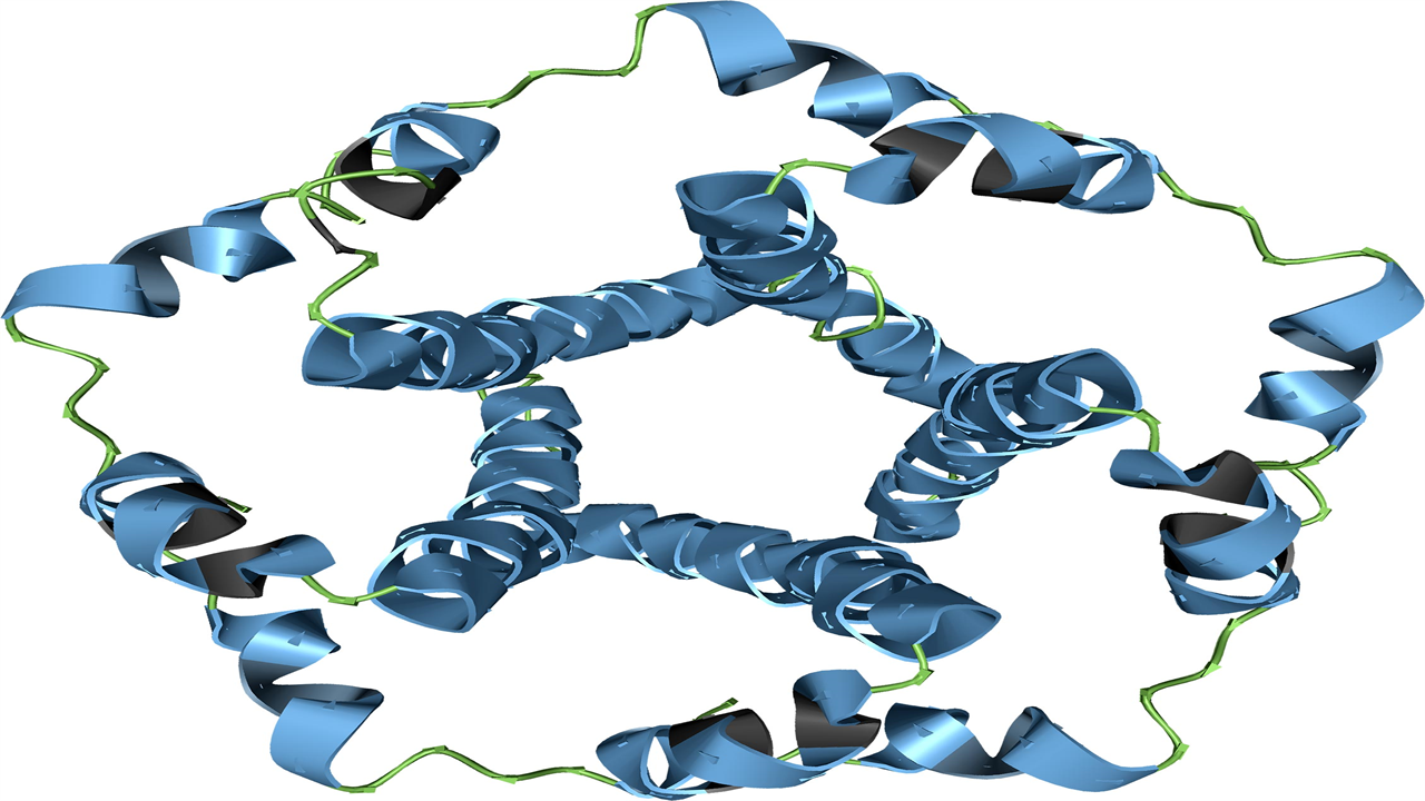
SARS-CoV-2 RNA Synthesis Complex Modeled in Aquariums. Credit: Garvan Institute of Medical Research
The team’s analysis revealed three coronavirus proteins (NSP3, NSP13 and NSP16) that “mimic” human proteins, which the researchers believe may help the virus hide better from the human immune system and contribute to the variation in COVID-19. outcomes.
The modeling also revealed five coronavirus proteins (NSP1, NSP3, spike glycoprotein, envelope protein and ORF9b protein) that the researchers say “hijack” or disrupt processes in human cells, allowing the virus to control its life cycle. completes and spreads. to other cells.
“Furthermore, we found eight coronavirus proteins that self-assemble with each other – analyzing how they assembled has yielded new insights into how the virus replicates its genome. However, after accounting for overlaps, there are still 14 proteins that we believe play a key role in infection, but have no structural evidence of interaction with other viral or human proteins,” said Professor O’Donoghue.
SARS-CoV-2 Spike Glycoprotein and ACE2 Protein Modeled in Aquariums. Credit: Garvan Institute of Medical Research
“To make all these insights and data more accessible to researchers, we have devised a new visualization method, a structural coverage map. The map shows what we know about SARS-CoV-2 and what remains to be discovered – it also helps scientists find and use 3D models to explore specific research questions.”
viral monitoring
The team’s analysis reveals opportunities for further research. “Much of the coronavirus research to date has focused on the spike glycoprotein, the main target for current vaccines. This protein will remain an important target, but it is also important that we broaden our focus to other potential targets and better understand the full viral life cycle,” said Professor O’Donoghue.
He adds that the Aquaria-COVID resource could help researchers more easily explore how new variants of coronavirus differ — and, critically, how to better target them with vaccines and treatments.
“The longer the virus circulates, the more likely it is to mutate and form new variants, such as the Delta strain,” said Professor O’Donoghue. “Our resource will help researchers understand how new strains of the virus differ from one another — a piece of the puzzle that we hope will help deal with new variants as they arise.”
Reference: “SARS-CoV-2 Structural Coverage Map Reveals Viral Protein Assembly, Mimicry, and Hijacking Mechanisms” September 14, 2021, Molecular Systems Biology.
DOI: 10.15252/msb.202010079
This research was supported by the Sony Foundation Australia, Tour de Cure Australia, the Wellcome Trust, Biotechnology and Biological Sciences Research Council and the Bundesministerium für Bildung und Forschung (BMBF).
This project was a collaboration between the Garvan Institute of Medical Research, CSIRO Data61, UNSW Sydney, Weihenstephan-Triesdorf University of Applied Sciences, Technical University of Munich, The University of Dundee and University College London.
Professor O’Donoghue is a Conjoint Professor, School of Biotechnology and Biomolecular Sciences (BABS), UNSW Sydney and a Visiting Scientist, Commonwealth Scientific and Industrial Research Organization (CSIRO) Data61, Australia’s national scientific agency.